Our lab focuses on two major themes – 1) tissue regeneration in axolotl and 2) next-generation methodologies
- Tissue regeneration in axolotl – Axolotls are the premier model to study tissue regeneration and rightly so because they can regenerate many different body parts. They can regenerate their legs, tail, and many different internal organs such as the heart, liver, lung, kidney, spinal cord, and even brain. Our lab is interested in, if they can, why can’t we? Further, they do so without fibrotic scars. Contrary, mammals are poor at tissue regeneration. Instead, injury in mammals lead to fibrosis. It is our hope that by studying mechanisms of tissue regeneration, one day we will be able to solve the problem of fibrosis.
-
Next-generation methodologies – Our lab firmly believes that we are asking the same biological questions that scientists have been asking for the last 200 years. For example, how does an embryo develop into an organism? or why do some organisms regenerate whereas others don’t? While the questions remain same, methodologies that are used to answer these questions have evolved. Our lab is constantly working to improve and implement next generation methodologies with a hope that these methodologies will help us unravel some of the biggest mysteries of biology.
Research Topics
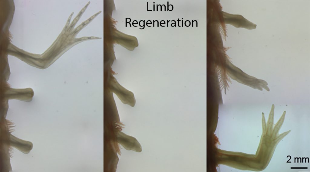
Limb regeneration
The limb is a complex structure that consists of numerous tissue types such as epidermis, bones, muscles, fibroblasts, nerves, vasculature and immune cells. Upon limb amputation all these cell types coordinate with each other and carry out an extraordinary feat of restoring exactly the lost portion. How do the cells even know where the amputation was made? How do they know when to stop regenerating? How do they form an exact replica with all the proper skeletal elements? These are just some of the questions that keep us busy.
Tail regeneration
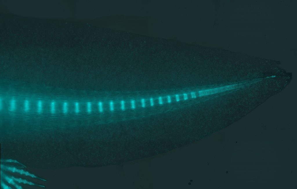
Axolotl is one of the few organisms that can regenerate their primary body axis including spinal cord. During embryonic development, an array of myotomes and vertebrae is formed through a segmentation process called somitogenesis. Upon tail amputation axolotls also recreate new segments, each containing new muscles and vertebrae. However, these segments originate from a mature tissue and in the absence of somites. Using state of the art technologies, we are addressing questions such as; what is the cellular source of the tail blastema and what are the underlying molecular mechanisms of tail regeneration.
Tissue metamorphosis
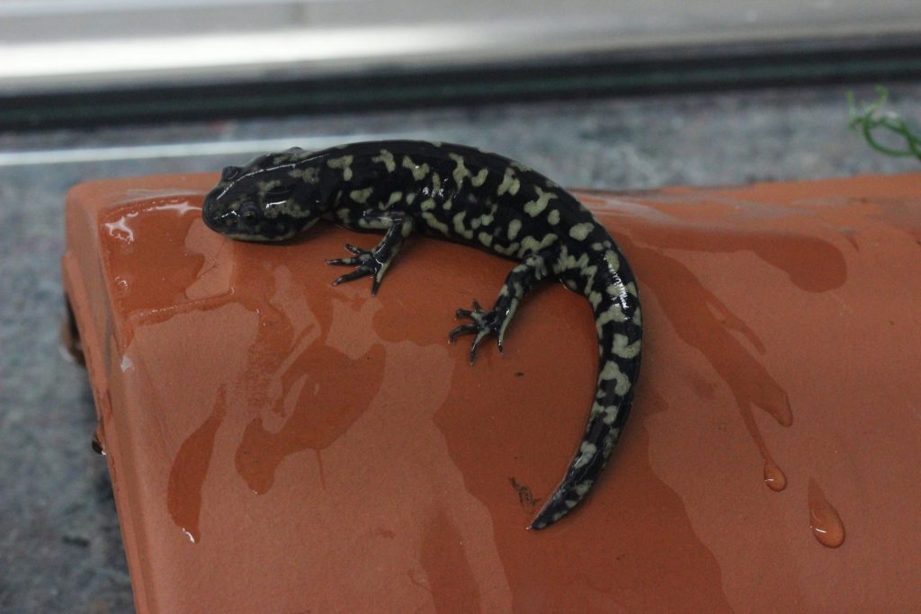
Axolotls are full of wonders. Although they spend most of their life in neoteny, in the lab they are capable of metamorphosis. A single exposure to L-thyroxine transforms axolotl body – they retract their gills and start breathing with their lungs. During metamorphosis they shed their skin and the emerging skin is more compatible with the terrestrial habitat. They lose their fin and their tail rounds up. Interestingly, they can still display tissue regeneration ability, although there is a small decline in the rate and fidelity. We want to understand the cellular and molecular basis for metamorphosis and its implication on tissue regeneration.

Transgenic methodologies
Genetic manipulations have completely revolutionized biological science. While transgenic methodologies were widely deployed in mice, zebrafish, and drosophila since the 90s, the axolotl lagged behind. We have spent a lot of time refining and improving these methodologies, which now allow us to develop I-SceI transgenesis, CRISPR-mediated knock-in and knockout, tissue-specific fluorescent reporters, and inducible Cre-loxP methodologies in axolotl. While much is accomplished, there is still a lot of scope for improvement. We are continuously working to improve these methods.
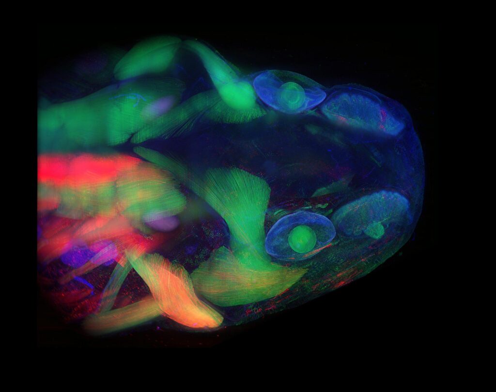
Tissue clearing
How wonderful it would be, if we do not have to deal with the opacity of tissues and organs. Just a thought that we can see through regenerating tissue is breathtaking. While nature has made few transparent organisms, most models are non-transparent. However, by understanding the principles of light behaviors and its interaction with various substances/chemicals, we can achieve high level of transparency in fixed tissues. Our lab is constantly investigating these principles and leveraging it to perform whole mount imaging to perform snapshots of regeneration.
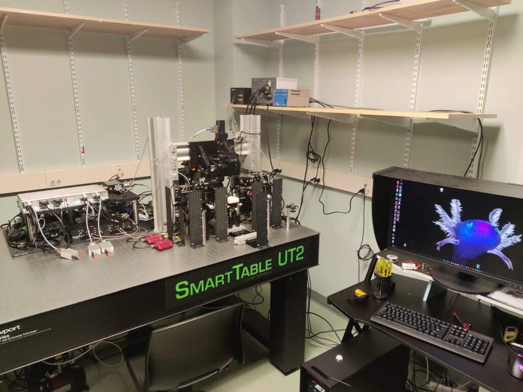
mesoSPIM microscopy
With support from the developers of the mesoSPIM (please see www.mesospim.org for more information), we have built a next-generation mesoSPIM (mesoscale selective plane illumination microscopy) microscope at MDIBL. The funding to build this microscope was generously supported by NIGMS. The mesoSPIM microscope enables imaging of multi-cm scale samples at a subcellular resolution. At the time of its construction, this was the fourth mesoSPIM microscope on the American continent. It was developed in December 2022 and placed under the care of MDIBL-LMF which has made it accessible to other national and international researchers.